Pregnancy is a special journey, and modern technology has made it easier for parents to see and understand their baby’s development. One of the most exciting advancements is ultrasound imaging. Many parents today are curious about 3D vs 4D ultrasound and how these two methods differ.
While both options give a clearer picture than traditional 2D scans, they serve slightly different purposes. In this article, we will explain 3D vs 4D ultrasound in simple terms so you can understand which one may be right for you.
What Is an Ultrasound in Pregnancy?
Ultrasound is a safe and painless imaging test that uses sound waves to create pictures of your baby inside the womb. Doctors use it to monitor growth, check health, and detect any concerns early.
As technology has improved, ultrasound has moved from flat images (2D) to more advanced options like 3D ultrasound and 4D ultrasound. This is where the comparison of 3D vs 4D ultrasound becomes important.
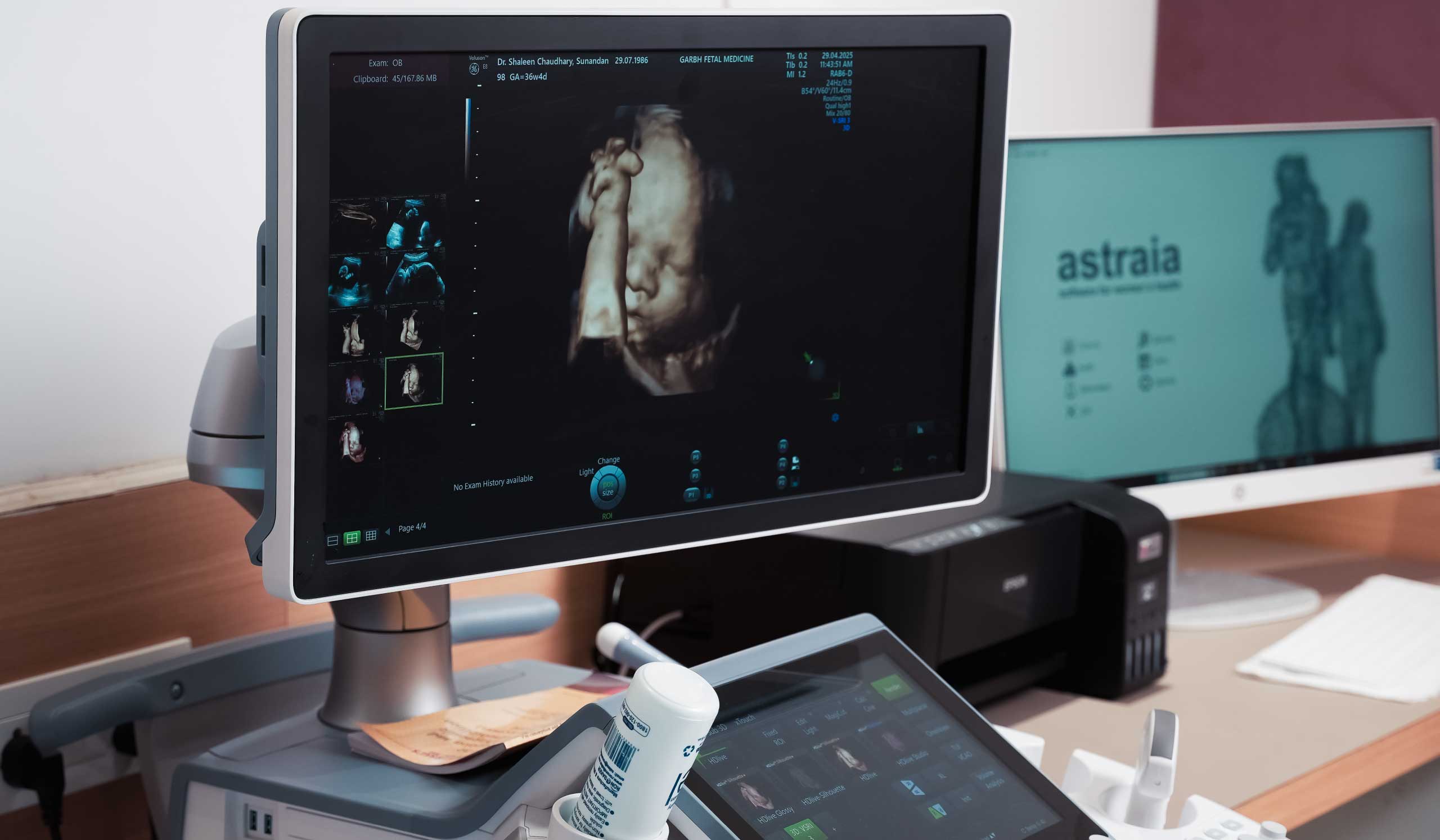
What Is a 3D Ultrasound?
A 3D ultrasound creates a three-dimensional image of your baby. Instead of flat pictures, it combines multiple images to show depth, shape, and structure.
With a 3D ultrasound, you can clearly see your baby’s face, hands, and other features. It gives a more realistic view compared to standard scans. Many doctors use this method to examine physical development and check for visible abnormalities.
When discussing 3D vs 4D ultrasound, it’s important to know that 3D scans provide still images, not movement.
What Is a 4D Ultrasound?
A 4D ultrasound takes things one step further. It adds motion to 3D imaging, allowing you to see real-time video of your baby.
This means you can watch your baby move, stretch, yawn, or even smile. A 4D ultrasound provides a live experience, making it more engaging for parents.
In the debate of 3D vs 4D ultrasound, this real-time feature is what makes 4D scans stand out.
3D vs 4D Ultrasound: Key Differences
Let’s break down the main differences between 3D vs 4D ultrasound in a simple and clear way.
1. Real-Time Imaging
The biggest difference in 3D vs 4D ultrasound is how images are captured.
A 3D ultrasound produces still images that show your baby’s structure. These images are detailed but do not show movement.
A 4D ultrasound, on the other hand, shows live motion. You can watch your baby move in real time, which gives a better understanding of behavior and activity.
2. Image Depth and Detail
When comparing 3D vs 4D ultrasound, both provide depth and clarity.
A 3D ultrasound shows detailed body structures, helping doctors examine facial features and body parts more clearly.
A 4D ultrasound includes all the benefits of 3D but adds motion, making it easier to understand how the baby is positioned and moving.
3. Viewing Facial Features
Another interesting point in 3D vs 4D ultrasound is how the baby’s face is seen.
With a 3D ultrasound, you can get a clear still image of your baby’s face. Many parents love this because it gives them the first “photo” of their child.
With a 4D ultrasound, you can see facial expressions as they happen. Your baby might blink, yawn, or move their mouth, which makes the experience more emotional and exciting.
4. Monitoring Baby’s Movement
Movement is another key factor in 3D vs 4D ultrasound.
A 3D ultrasound cannot show movement since it captures still images.
A 4D ultrasound allows doctors to observe how your baby moves. This helps in understanding muscle development and overall well-being.
5. Purpose and Use
When looking at 3D vs 4D ultrasound, both have medical and personal uses.
A 3D ultrasound is often used for checking physical development and identifying visible abnormalities.
A 4D ultrasound is useful for both medical observation and bonding. It gives parents a chance to see their baby in action.
6. Cost and Availability
Cost is another consideration in 3D vs 4D ultrasound.
A 3D ultrasound is usually more affordable and widely available.
A 4D ultrasound may cost more because of advanced technology and may not be available in all clinics.
When Do You Need a 3D or 4D Scan?
Doctors recommend ultrasound scans at different stages of pregnancy. While standard 2D scans are routine, 3D and 4D scan options may be suggested in certain cases.
You may need a 3D and 4D scan for:
- Checking baby’s growth and development
- Looking for physical abnormalities
- Observing facial structures
- Monitoring fetal movement
- Enhancing bonding with your baby
In some clinics, parents also choose a 3D and 4D scan for keepsake images or videos.
Benefits of 3D and 4D Ultrasounds
Understanding the benefits helps in choosing between 3D vs 4D ultrasound.
Both types offer clearer and more detailed images than traditional scans. They help doctors detect certain conditions earlier and give parents reassurance.
A 3D ultrasound is especially helpful for examining structural issues like cleft lip.
A 4D ultrasound provides additional insight into how the baby moves, which can be useful for monitoring development.
Are 3D and 4D Ultrasounds Safe?
Safety is a common concern when discussing 3D vs 4D ultrasound.
Both 3D ultrasound and 4D ultrasound use the same sound wave technology as standard ultrasounds. They are considered safe when performed by trained professionals.
However, they should only be done when medically necessary or recommended by your doctor.
Choosing Between 3D vs 4D Ultrasound
When deciding between 3D vs 4D ultrasound, your choice depends on your needs.
If you want detailed still images for medical evaluation, a 3D ultrasound may be enough.
If you want to see your baby moving and enjoy a more interactive experience, a 4D ultrasound may be the better option.
In many cases, doctors may recommend the option that best suits your pregnancy condition.
FAQs
What is the difference between 3D and 4D ultrasound in pregnancy?
Ans.The main difference in 3D vs 4D ultrasound is that 3D creates still images, while 4D shows real-time movement of the baby.
When is the best time to get a 3D or 4D ultrasound during pregnancy?
Ans.The ideal time for a 3D and 4D scan is usually between 26 and 32 weeks of pregnancy when the baby’s features are more developed.
Can 3D or 4D ultrasound detect birth defects?
Ans. Yes, both 3D ultrasound and 4D ultrasound can help detect certain physical abnormalities, especially those related to facial structure.
Which scan is done first: NT scan or anomaly scan?
Ans. In the comparison of NT scan vs anomaly scan, the NT scan is done first, followed by the anomaly scan later in pregnancy.
Why do doctors recommend both NT scan and anomaly scan?
Ans. Doctors recommend both scans because NT scan vs anomaly scan highlights different aspects of fetal health, one screens for chromosomal risks, while the other detects structural abnormalities.
Why do doctors recommend a 3D or 4D ultrasound?
Ans.Doctors may suggest a 3D and 4D scan to get a clearer view of the baby’s anatomy and movements for better diagnosis.
How long does a 3D or 4D ultrasound scan take?
Ans.A 3D and 4D scan usually takes around 20 to 45 minutes, depending on the purpose and baby’s position.
Which is better: 3D or 4D ultrasound?
Ans.In 3D vs 4D ultrasound, neither is strictly better. 3D is good for still images, while 4D is better for viewing movement.
Are 3D and 4D ultrasounds necessary in pregnancy?
Ans.No, they are not always necessary. Standard scans are usually enough, but 3D and 4D scan options can provide additional information when needed.
Where can you get a 3D or 4D ultrasound during pregnancy?
Ans.You can get a 3D and 4D scan at specialized clinic like The Garbh that offer advanced imaging services.
Conclusion
Understanding 3D vs 4D ultrasound can help you make better decisions during pregnancy. Both options offer advanced imaging and valuable insights into your baby’s development.
A 3D ultrasound gives clear, detailed images, while a 4D ultrasound brings those images to life with movement. Choosing between them depends on your medical needs and personal preference.
Always consult your doctor before opting for any scan to ensure the best care for you and your baby.

